Perth Animal Eye Hospital Diagnostic Facilities
Determining the cause of an eye problem can be difficult, so having the best available diagnostic equipment is essential. At Perth Animal Eye Hospital we have the most up to date diagnostic facilities available, together with the expertise to correctly identify the cause of the problem, and enabling correct therapeutic strategies to be formulated, thus helping to resolve the problem as quickly as possible
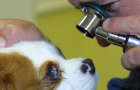
Direct ophthalmoscopy
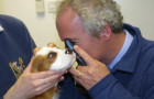
The direct ophthalmoscope is used to examine the clarity of the eye, checking for any abnormality that blocks the passage of light in the eye. This instrument is also used to examine the back of the eye in great detail, helping to diagnose diseases of the retina or optic nerve.
Indirect ophthalmoscopy
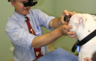
Indirect opthalmoscopy is used to examine the back of the eye. This technique gives a wide field of view which improves the ability to detect subtle or focal changes.
Slit lamp biomicroscopy
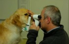
The slit lamp biomicroscope is a highly specialised tool used to look at the eye in microscopic detail. This instrument can localise minute abnormalities such as fine hairs or foreign bodies that might irritate the eye, help determine the type, depth and cause of corneal ulcers, check for inflammation inside the eye, and help to diagnose and localise cataracts.
Tonometry
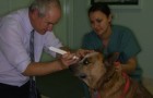
Tonometry is the measurement of intraocular pressure. This is important in the diagnosis of glaucoma and inflammation inside the eye. Tonometry at Perth Animal Eye Hospital is performed using very advanced equipment (Tonopen and TonoVet tonometers), providing accurate assessment of the pressure inside the eye.
Gonioscopy
This is a technique used to examine the fluid outflow pathways in the eye and helps to determine the cause of glaucoma. Glaucoma is a devastating disease which often leads to blindness and even removal of the eye if incorrect treatments are employed. Gonioscopy is an essential part of glaucoma diagnosis as it can help identify conditions that might be causing high pressure in an eye, but often more importantly it can help to determine if the other eye is at risk, allowing prophylactic measures to be taken to prevent blindness in the second eye. There are many causes for glaucoma, and treating the wrong form of glaucoma with the wrong medication will make the eye much worse. Gonioscopy allows for the proper medication to be chosen or may demonstrate that surgery provides the best outome for the eye and vision.
Ultrasound
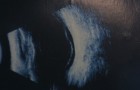
Ultrasound examination can be used to hep to identify abnormalities inside the eye when the eye is too cloudy to examine directly. It can be useful to diagnose and localise tumours inside the eye, lens problems that might cause glaucoma, and retinal detachments. It can also be used to examine problems behind the eye, such as orbital inflammation or tumours. At Perth Veterinary Ophthalmology the ultrasound machine is an Ultrasound Biomicroscope (UBM) capable of providing very high magnification and detailed images.
Electroretinography (ERG)
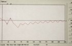
An ERG machine helps to record electrical activity in the retina and can be used to help determine the cause of sudden onset blindness, or to ensure that the retina is functioning normally prior to cataract surgery.
Blood pressure measurement
High blood pressure can lead to blindness haemorrhage inside the eye and also can cause retinal detachments. It can also predispose animals to kidney disease, heart disease and brain disease. High blood pressure can occur in both both cats and dogs, but cats are particularly susceptible. At Perth Veterinary Opthalmology we can measure blood pressure to test for high blood pressure and monitor response to treatment.